


Comprehensive Brain Tumor Program
Learn about our leading-edge brain tumor treatment options. We’ll custom tailor our nationally recognized program to suit your needs.
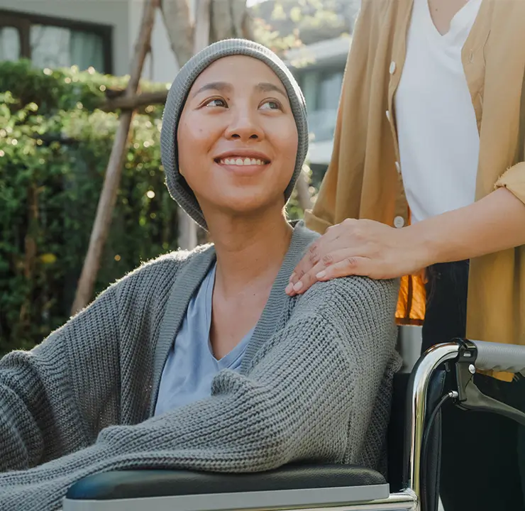
When you partner with us, know that our care ranges from groundbreaking treatments to help with navigating basic daily tasks.
Our approach to comprehensive brain tumor care
We understand the stress that comes with a new and frightening brain tumor diagnosis. Because many brain tumors grow so fast, we’ll help you make thoughtful yet rapid treatment decisions. That way you can return to living life as soon as possible.
In the Brain Tumor Program, our top priority is to give you individual attention. We offer compassionate care blended with full access to highly advanced treatment approaches. If you qualify, you may be able to participate in the most promising clinical trials that exist nationally and internationally.
How you’ll work with us
Your health journey is completely unique. But these are the general steps you can expect to take.
- Expedited clinic visits – you’ll meet with neuro-oncology, neurosurgery and radiation therapy specialists in one convenient visit.
- Imaging, biopsy and diagnosis – benefit from precise imaging techniques, tissue samples and a pinpoint accurate diagnosis.
- Discuss customized treatment options with your doctor.
- Get education and support – help is available.
We’ll make sure you understand every detail and will help you solve practical obstacles getting in the way of your treatments.
Second opinions
If you need a second opinion, we are a powerful, nationally recognized resource.
We promise a quick response. If we are not immediately available, one of our physicians will call you back within 24 hours. With insurance approval, you can make an appointment within 48 hours.
If you’re not local to Orange County, California and need a second opinion, contact us to make a virtual appointment.
Why choose UCI Health for the Comprehensive Brain Tumor Program?
Our Comprehensive Brain Tumor Program is part of the UCI Health Chao Family Comprehensive Cancer Center
We are the only program in Orange County affiliated with a National Cancer Institute-designated comprehensive cancer center. We earned that recognition from conducting innovative research and hundreds of clinical trials that could accelerate your recovery.
As a UCI Health brain tumor patient, we’ll automatically consider you for trials. You could get new, experimental treatments that are not widely available anywhere else. Some of our groundbreaking research has changed the way brain tumor care is delivered at a national level.
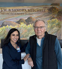
Our tumor board consists of 40 experts who meet weekly
They consult with each other regularly to create personalized treatments for our most complex cases.
We are also the only program in Orange County that offers you care from physicians with years of specialized (fellowship) training in brain tumors.
We know how and when to make the right recommendation based on years of evaluating hundreds of patients
The right recommendation isn’t always based on the size of your tumor. We prescribe treatments from a deep well of expertise and ongoing multidisciplinary discussions. Our Brain Tumor Program doctors only treat brain tumors and that high volume of daily experience is exceptionally valuable.
Ultimately, we use that expertise to deliver what works best for you.
We educate our physicians working in general neurology, primary care and internal medicine about the early signs and symptoms of brain tumors
We increase our clinicians’ awareness about when it’s appropriate to order a brain CT or a brain MRI. For example, many of our older patients with brain tumors have cognitive difficulties that look very similar to Alzheimer’s disorder. If a neurologist suspects Alzheimer’s, in the diagnosis of exclusion, it’s very important that they get a brain scan first before making an Alzheimer’s diagnosis.
Looking for more options?
View all clinicians
Our brain tumor support is truly comprehensive
A brain tumor diagnosis isn’t just physical, it affects you emotionally and spiritually too. We offer multidisciplinary care across all three dimensions to help you reconnect with life.
Reach out and make an appointment with a brain tumor expert. Call 714-456-8000.

Find a cancer clinical trial
Talk to your doctor to see if a cancer clinical trial is right for you.
We welcome referrals from community physicians
To refer a patient to our program, call 714-456-8000 or fill out an online request form. You may also email us to make an appointment.
One of our brain tumor physicians will return your call within 24 hours. We can see your patient in our offices within 48 hours after insurance approval.
Featured Blog Posts
Novel ALS therapy a 'game-changer' for artist

Living well during cancer treatment