





Ophthalmology | Gavin Herbert Eye Institute
Our UCI Health Gavin Herbert Eye Institute specialists have one mission: deliver outstanding patient care.
Whether you need a routine eye exam, LASIK surgery or care for complex vision problems, our internationally renowned ophthalmologists can help you.
As Orange County's only academic eye center, our surgeons and scientists not only provide you with the most advanced eye care, but they also offer access to leading-edge clinical trials, sight-saving treatments and therapies for virtually any eye condition.
Why Choose Us
You can count on receiving the highest quality eye care at the UCI Health Gavin Herbert Eye Institute. Our nationally recognized specialists are at the forefront of research, developing and delivering the latest treatments for a wide range of eye conditions.
- Our team includes pioneers in the development and use of ophthalmic lasers and refractive surgery techniques. Their work has formed the foundations of LASIK surgery, the development of the blade-less IntraLase™ laser and the most advanced techniques for corneal transplants and intraocular lens implants.
- Our team has made critical advances in treating age-related macular degeneration, the leading cause of blindness in people over age 55.
- Our surgeons, stem cell researchers, geneticists, infectious disease specialists and engineers are working on technologies and treatments for diabetic retinopathy, retinitis pigmentosa, glaucoma, corneal disease and cataracts, as well as a vaccine to prevent eye and genital herpes infections.
- We also are international leaders in glaucoma implant surgery, having developed many of the techniques and technologies used around the world today.
Our institute's ophthalmologists bring this expertise and vision to the care they provide daily to patients with eye conditions that range from mild myopia to rare retinal and ocular disorders. Many of them appear regularly on the "Best Doctors in America®" list, a peer-review survey that represents the top 5 percent of specialists in the nation.
We see patients at the UCI Health Gavin Herbert Eye Institute on the university campus in Irvine, at UCI Medical Center's Pavilion 2 in Orange and at the UCI Health — Brea Outpatient Center.
Our clinicians
Cataracts, cornea, external disease and refractive
Soroosh Behshad, MD, MPH Marjan Farid, MD Sumit (Sam) Garg, MD Sanjay R. Kedhar, MD Olivia L. Lee, MD Matthew W. Wade, MD Junru Yan, MD
Comprehensive and cataracts
Alpa S. Patel, MD Farnoosh Tinoosh, MD
Glaucoma and cataracts
Austin R. Fox, MD Ken Y. Lin, MD, PhD Sameh Mosaed, MD Andrew K. Smith, MD Michael C. Yang, MD
Pediatric ophthalmology and strabismus
Charlotte Gore, MD Donny W. Suh, MD Stephen B. Prepas, MD
Neuro-ophthalmology
R. Wade Crow, MD Lilangi S. Ediriwickrema, MD Kimberly K. Gokoffski, MD, PhD Vivek R. Patel, MD Samuel J. Spiegel, MD
Oculoplastics/Orbit surgery
Lilangi S. Ediriwickrema, MD Seanna Grob, MD Jeremiah P. Tao, MD
Ocular oncology
Retina and vitreous
Andrew W. Browne, MD, PhD Baruch D. Kuppermann, MD, PhD Stephanie Y. Lu, MD Mitul C. Mehta, MD Kapil Mishra, MD Mohammad Riazi Esfahani, MD
Uveitis
Sanjay R. Kedhar, MD Olivia L. Lee, MD
Low vision
Pediatric optometry
Ophthalmology Conditions & Treatments
|
|
|
|
Register for our community lecture series
Our free lecture series highlights the latest advances in eye disease treatment while educating and empowering you and the community.

Participate in vision research and clinical trials
Our clinicians research the next generation of sight-saving devices, drugs and procedures. If you participate, you may have access to treatments not yet widely available.
Research
Gavin Herbert Eye Institute has assembled a unique, world-class group of scientists, engineers and physicians who conduct patient-oriented basic science and applied research on the next generation of sight-saving devices, drugs and procedures.
This internationally recognized team of investigators includes basic and clinical scientists who have placed UCI's School of Medicine in the top echelon of U.S. institutions receiving National Institutes of Health grants for vision research. Many are pioneers in the development of ophthalmic lasers and refractive surgical technologies. Others are at the forefront of stem cell treatments for such conditions as macular degeneration, retinitis pigmentosa and corneal disease. All are focused on developing the treatments of the future that will prevent vision loss.
Basic research studies currently include:
- Stem cells for retinal regeneration in macular degeneration and retinitis pigmentosa
- Regenerative medicine for retinal and corneal diseases
- Genetics of macular degeneration to improve treatment outcomes
- Safety testing of medications used in treating retinal disorders
- The role of environmental factors in retinal degeneration
- Imaging the optic nerve in glaucoma patients
- Development new therapies for glaucoma
- Understanding and preventing corneal scarring
- Understanding and preventing vision loss from herpes infections
Clinical research studies currently include:
- Clinical trials for diabetic macular edema, diabetic retinopathy and vitreous hemorrhage
- Clinical trials for both "wet" and "dry" age-related macular degeneration
- Clinical trials for retinal vein occlusion
- Clinical trials for vitreomacular traction
- Clinical trials for retinal detachment
- Clinical trials for imaging the optic nerve in patients with glaucoma and macular degeneration
- Clinical trials for imaging the retina and choroidal vasculature in retinal vein occlusion, diabetic retinopathy and age-related macular degeneration
- Evaluation of the Trabectome instrument in glaucoma treatment
- Development and testing of new corneal transplant techniques
Current clinical trials
FAQs
Frequently asked questions about ophthalmology at UCI Health.Myopia (nearsightedness)
Patients can see near objects clearly but things at a distance are blurred. Myopia usually results when the eye is too large or elongated, which causes light entering the eye to focus before reaching the retina.Hyperopia (farsightedness)
Patients have good distance vision but close-up objects appear blurred. Hyperopia is due to the eye being smaller than normal, causing light entering the eye from close-in objects to come into focus too far behind the retina.Astigmatism
This can impair both near and far vision. It occurs when the cornea and eye lens is oval in shape instead of round, which lets light focus on several points on the retina instead of one, causing blurring. Astigmatism often occurs with myopia or hyperopia.Presbyopia (aging eyes)
Most patients begin to experience “old-eyes” in their 40s, a function of natural aging as the eye lens hardens and loses the flexibility to focus on close-up objects.A cataract is a clouding of the transparent lens inside the eye behind the colored iris that prevents light from reaching the retina. It results in blurred vision, glare and halos around objects. There are three types of cataracts:
- Nuclear cataracts form in the nucleus or center of the lens, usually as a patient ages.
- Cortical cataracts form in the cortex, outside the eye lens. Over time, this cataract extends from the outside to the inside of the lens. They are common in diabetic patients.
- Subcapsular cataracts form at the back of the lens. This type of cataract is common in diabetics, people taking steroid medication and those who are farsighted or have retinitis pigmentosa.
This microsurgery, involving tiny incisions, is performed in an outpatient ambulatory surgery center. In most cases, the procedure is completed painlessly in less than 20 minutes. Most patients find their vision improved immediately after the surgery.
Macular degeneration, or age-related macular degeneration (AMD), is a disease that gradually destroys the macula, the part of the eye that is responsible for central visual acuity.
A leading cause of vision loss in Americans age 60 and older, AMD causes no pain and, in some cases, advances so slowly that people notice little change. In other patients, the disease progresses quickly and may lead to a loss of vision in both eyes. It occurs in two forms:
- In dry AMD, the macula’s light-sensitive cells break down slowly, leading to a buildup of waste product and causing a gradual blurring of central vision. Over time, as less of the macula functions, central vision is lost in the affected eye.
- In wet AMD, abnormal blood vessels behind the retina start to grow under the macula. These vessels tend to be fragile and often leak blood and fluid, which pushes the macula from its position at the back of the eye and results in rapid loss of central vision.
An early symptom of wet AMD is that straight lines appear wavy. In dry AMD, the most common symptom is slightly blurred vision, difficulty recognizing faces, reading and performing other tasks. The central vision is affected.
To treat AMD, your physician may recommend ultraviolet sunglasses (especially the blue blocker type) and dietary supplements (vitamins A, C, E, Lutein, Omega 3 fatty acid, and the mineral zinc) to slow down the progression of the dry AMD.
Anti-VEGF (vascular endothelium growth factor) therapy is the main therapeutic agent used to treat wet AMD.
Occasionally photodynamic therapy, vision rehabilitation or laser treatments are also used.
Untreated, glaucoma is a leading cause of irreversible blindness in the United States, affecting more than four million Americans. With early detection, consistent monitoring, and prompt treatment, medicines (and surgery in refractory cases) can arrest the progression of glaucoma and preserve vision.
Generally, as we age, the skin thins out, tissue stretches and sags and the soft tissue loses volume. This often becomes apparent first in the eyelids.
Heavy upper eyelids (ptosis) may interfere with vision and can be fixed surgically. Loss of elasticity of the skin and sagging, baggy upper or lower eyelids also can be treated surgically.
Eyelids also can turn in towards the eye (entropion) or turn away from the eye (ectropion). They also may develop a range of growths from benign warts and styes to malignant lesions, such as basal cell carcinoma. These may need removal and reconstruction.
Questions? Contact the UCI Health Refractive Surgery LASIK Center at 949-824-9970.
Featured News Stories
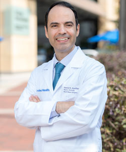
Understanding how the brain interprets sounds can help address ear ringing
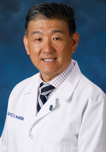
Identifying the causes of nearsightedness
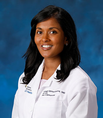
Reconstructive eye surgeon chosen for elite vision fellowship
Featured Blog Posts

A mother's legacy: Supporting vision research
The care Renee Horner's mother received at the Gavin Herbert Eye Institute launched a family tradition of giving.
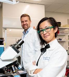
Eye researchers at work on groundbreaking whole-eye transplant
UCI Health neuro-ophthalmologist Dr. Kimberly Gokoffski and vision scientist Frans Vinberg are part of a U.S. effort to make eye transplants possible.

Author credits eye specialist for lifesaving cancer diagnosis
Upcoming Events
7-8 p.m.
Gavin Herbert Eye Institute Community Lecture Series
Attend the virtual UCI Health Gavin Herbert Eye Institute Community Lecture Series to learn about the symptoms, causes and treatments for eye-related conditions. RSVP is required. Register today via Zoom. For more information, email ophthalmology@uci.edu.